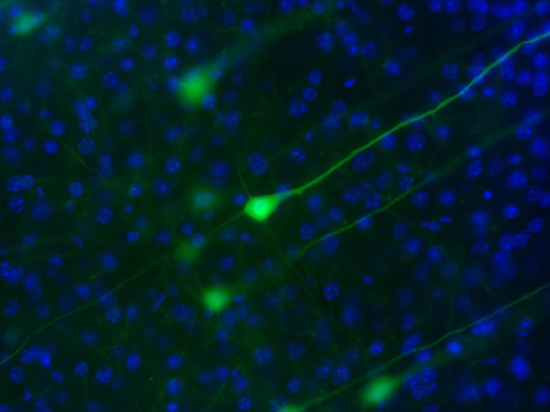
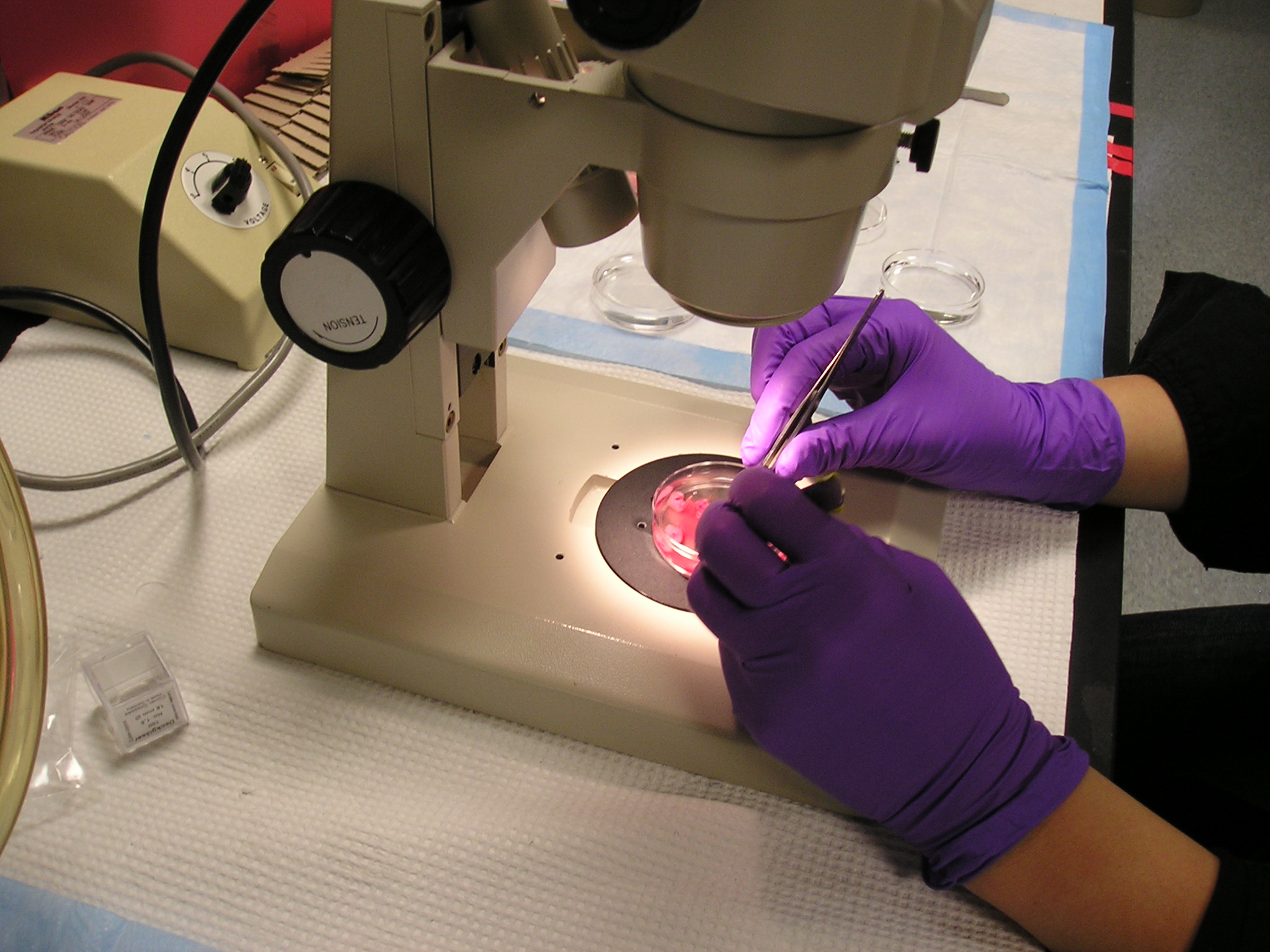
UPPER MOTOR NEURONS (BRAIN) ------- DISSECTING RODENT SPINAL CORDS ----- LOWER MOTOR NEURONS (SPINAL CORD) ---- GLIAL-NEURONAL MIXED CULTURES
Why do upper and lower
motor neurons degenerate in ALS? Our ability to move our
arms and legs depends on the integrity of our spinal motor neurons, which
relay electrical signals from our brain to our muscles. When these motor
neurons die, as can occur in injury or in a variety of degenerative diseases
that affect infants and children (spinal muscular atrophies) or adults (ALS)
irreversible paralysis or even death may ensue. Cortical upper motor neurons
extend their axons great distances from the brain down to the spinal cord. Spinal lower motor neurons then take the signal out to your muscles. In fact both of these cells are among the biggest cells in your body, and you have these cells for your entire life. For unknown
reasons in ALS, these select group of neurons in thebrain and spinal cord degenerate and die. A number of transgenic
animal models for ALS exist based on superoxide dismutase (SOD1) and TDP-43 gene
mutations found in a portion of familial ALS patients. These animals display
similar clinical symptoms and histopathology to that found in human motor
neuron disease. Both humans and the animal models show that there are abnormalities not only in the motor neurons but also in the surrounds other cells such as interneurons, astrocytes, oligodendrocytes, and microglia that communicate continuously with the motor neurons. Thus, it is now possible and crucial to understand the signaling
mechanisms that normally promote upper and lower motor neuron survival and
how these processes go awry in motor neuron diseases.
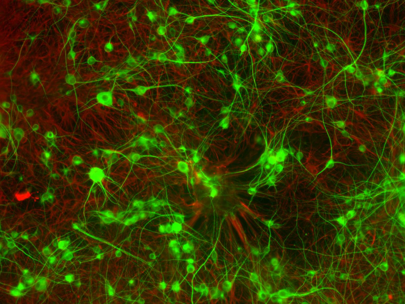
Our lab is focusing on understanding the basic cellular
and molecular pathogenesis of ALS by studying defined neuronal and glial
cells in culture that may be important in the degeneration and survival
of motor neurons. In addition, we use rodent animal models of the disease
to see how these cells and molecules interact in a complex organism that
gets an ALS-like disease. Finally, we are studying epidemiology and human
genetic polymorphisms of ALS in the human disease and working with teams of scientists and clinicians in clinical trials
with our patients.
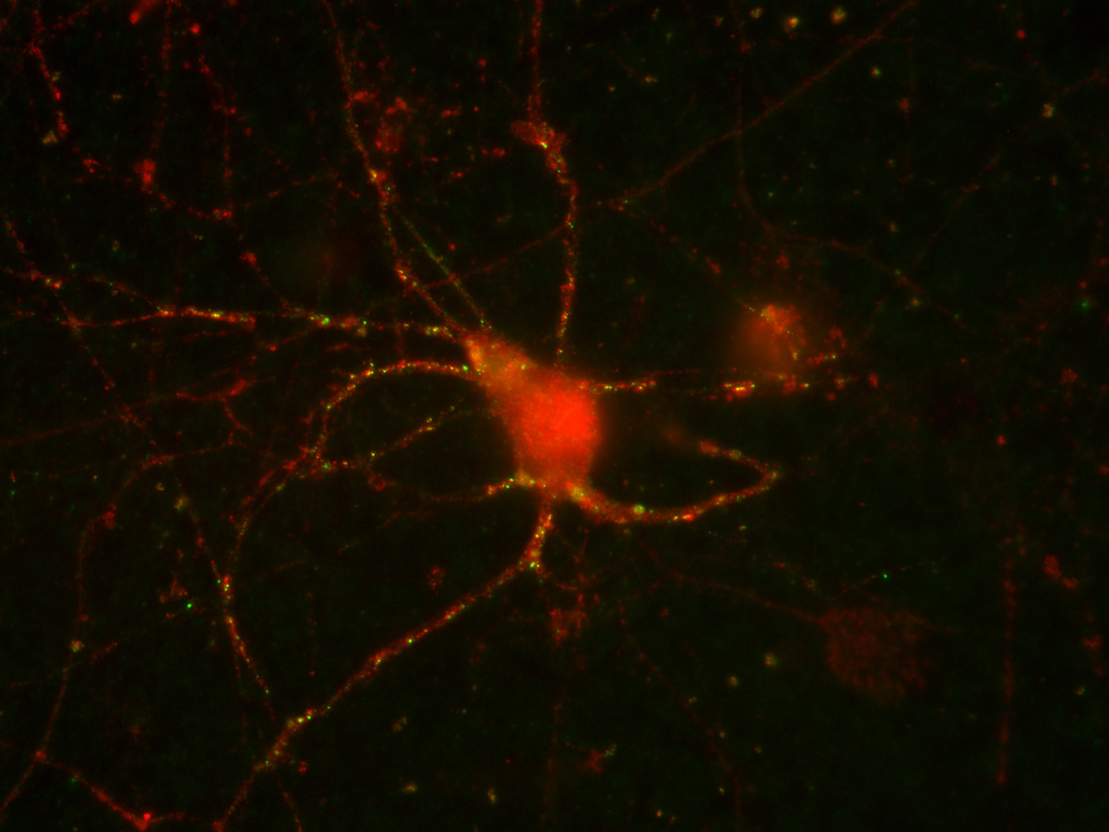
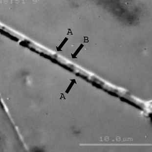
Axonal transport and mitochondrial dysfunction have been implicated in the pathogenesis of ALS.
Panel 1.Representative DIC computer enhanced image
of MN with vesicles (A) and mitochondria (B) moving in antero- and retrograde
directions. Panel 2: Fluorescently labelled mitochondria moving in MN processes. Click on the images to view movies of axonal transport in progress.
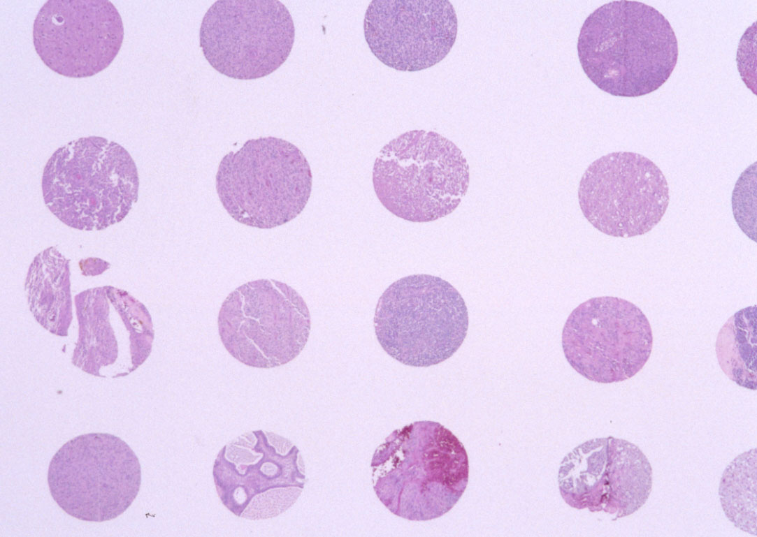
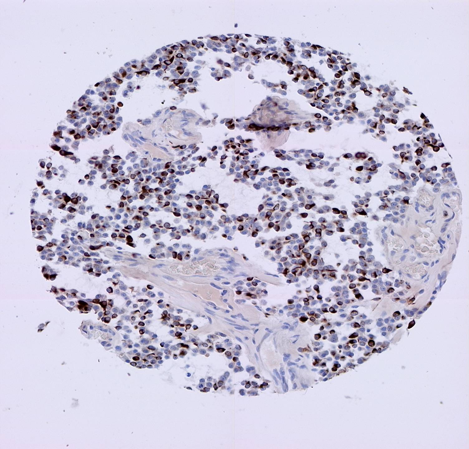
Tissue Arrays allow controlled sampling of dozens
to hundreds of tissue samples on a single slide. Our laboratory is interested
in using this technique to study brain, spinal cord, and nerve tumors in conjunction with whole-slide imaging technology.
Fluorescence assisted Resection of CNS Tumors
Novel methods of fluorescently labelling brain tumors to assist neurosurgeons with more complete resections. Our lab in collaboration with folks at Dartmouth Medical School and the Thayer School of Engineering is studying the pathology and validating the techniques for this tranformational technique in neurosurgery.
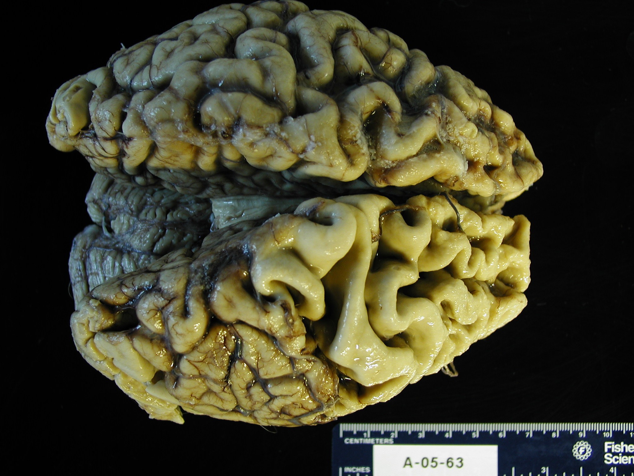
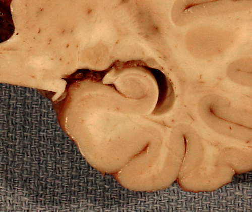
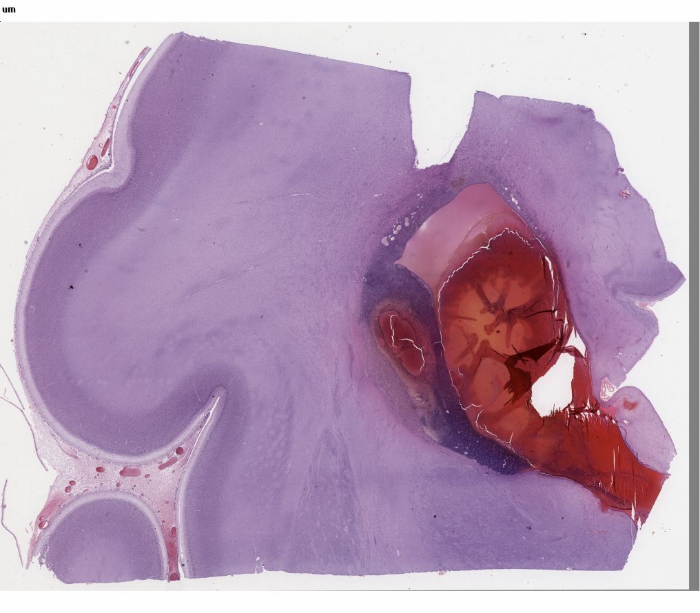
GEORGETOWN BRAIN BANK
FRONTO-TEMPORAL LOBAR DEGENERATION - NORMAL HIPPOCAMPUS - GERMINAL MATRIX HEMORRHAGE
The medical autopsy remains the “gold standard” for diagnosing and classifying neurological diseases. Autopsies also allow (when permitted) collection of tissues and biofluids for research into the pathology of the specific diseases. However, reimbursement for autopsies by the federal government even at academic medical centers is no longer being meaningfully funded. As a result autopsy rates have dramatically decreased in recent years. This is occurring at a time when small and large scale clinical trials for new neurotherapeutics desperately need to have established diagnosis during the trial and confirmed upon autopsy and at a time when the genetics of diseases need to be correlated with pathological and clinical findings.
We have established the Georgetown Brain Bank (GBB) to serve three key constituents of our academic medical center: our patients, our students, and our researchers. Families and the clinical teams caring for our patients with neurological diseases will benefit from a careful neuropathological examination to provide diagnoses and context to patients’ illness and valuable information for descendents. Medical students, neuroscience graduate students, and housestaff will benefit from educational sessions on gross and microscopic neuropathology. The research community will have access to well characterized tissues/fluids to allow for translational research projects and a collaborative neuropathologist to assist them. Clinical researchers will be able to confirm diagnoses crucial to understanding results of their trial and potential adverse effects.
COLLABORATIONS:
Our lab is also committed to providing expertise in neuropathology and
neuroscience techniques to the Georgetown neuroscience community and beyond,
and we have established collaborations with
individuals from Pathology, Neurology, Neurosurgery,
LCCC.